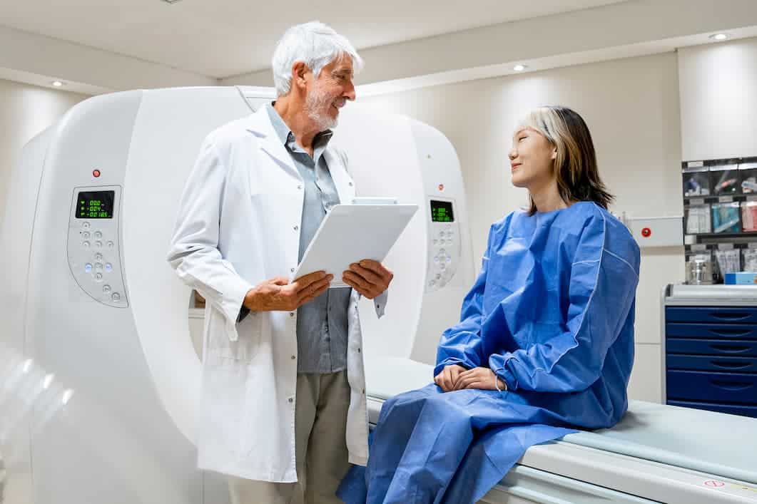
Radiologists in Montclair, New Jersey
The imaging department at Mountainside Medical Center offers a full range of inpatient and outpatient radiology services, including diagnostic, therapeutic, and interventional procedures. Our imaging specialists are committed to providing the Montclair, New Jersey community with fast and efficient imaging services. By pairing compassionate care with advanced technology, we’re able to deliver accurate imaging services you can depend on. Our radiologists provide services like CT scans, PET scans, and ultrasounds, and we also offer diagnostic procedures for cardiovascular, musculoskeletal, and neurological conditions. No matter your needs, Mountainside Medical Center is prepared to meet them.

Our Diagnostic Imaging Services
Our imaging specialists are dedicated to providing outstanding care and accurate radiology services. We employ a team of radiologists certified American Board of Radiology, as well as skilled technicians and nurses to perform a variety of imaging procedures, including:
- Bone density scans (DXA)
- Computerized tomography (CT) scans
- Heartflow analysis
- Interventional radiology
- Laboratory services
- Magnetic resonance imaging (MRI)
- Mammography
- Nuclear medicine
- Positron emission tomography (PET) scans
- Ultrasound
- X-rays
Our outpatient imaging department is open daily, as well as in the evenings and on weekends, so that you can receive prompt and convenient imaging services whenever your physician prescribes a procedure. Same day appointments are available in many cases.
Free parking is provided for test and procedure patients in the parking lot conveniently located across the street from the Harries Pavilion entrance on Highland Avenue. Patients will receive a free parking token from the information desk upon return of their visitor badge. Free valet parking is also available.
Learn more about our tests
Bone density scans
Bone density scanning, also known as dual-energy, x-ray absorptiometry (DXA) or bone densitometry, is an enhanced form of x-ray technology used to measure bone mineral density (BMD) and loss of bone mass. DXA is most often performed on the lower spine and hips. However, the entire body is sometimes scanned. Body scans are painless, non-invasive procedures with great accuracy in detecting the presence and degree of lost bone mass.
We offer state-of-the-art bone scan testing to detect the presence of osteoporosis and other conditions that cause bone weakness. The Mountainside Bone Health Center can effectively diagnose and treat osteoporosis to prevent painful bone fractures and other medical problems that can result from accidents and falls due to bone fragility.
Fill out our bone density questionnaire
CT scans
CT scanning is a painless, non-invasive testing method widely used by doctors because it provides the most revealing images of internal organs, bones, soft tissues and blood vessels. Computed tomography (CT) combines special x-ray technology and sophisticated computer technology to create high-speed, cross-section images of the inner workings of the human body. The images are electronically reconstructed to create two and three-dimensional illustrations that can reveal a wide array of medical conditions.
Mountainside Medical Center is equipped with multiple CT scanners including a dual-source 256-slice Somotom. Our high speed, high-resolution Somatom CT scanner produces vivid images that are valuable tools for acute care, cardiology, oncology, neurology, and other areas of medicine. Enhanced resolution and image clarity ensures the accuracy of patient diagnoses, offering you better health outcomes and a more tailored treatment plan.
The Mountainside Medical Center radiology team is among a select group nationwide that includes two physicians with the highest, Level III certifications in cardiac CT angiography from the American College of Cardiology.
MRI
MRIs are used in the diagnosis of many, varied conditions and are often requested by physicians to obtain detailed information not already provided by other imaging technologies. Magnetic Resonance Imaging (MRI) employs strong electromagnets, radio frequency waves and powerful computers to generate two and three-dimensional images of the body’s organs, tissues and bones. Unlike x-rays and CT scans, MRI technology does not require the use of radiation.
Images are gathered using a large, tube-shaped magnet that creates a strong magnetic field around the patient. A radio frequency coil is placed over the body part that is to be imaged. The magnetic field and radio frequency waves alter the alignment of hydrogen protons within the body. Using signals emitted from the protons, computers reconstruct images of the body part to be studied.
Our areas of expertise include MRI angiography to examine the heart and other vessels and MR arthrography to examine joints including the knee and hip when standard x-rays are inconclusive. Our radiology team is also skilled in MRCP (magnetic resonance cholangiopancreatography), a non-invasive imaging technique used to visualize the biliary and pancreatic ducts to determine if gallstones are lodged in any of the ducts surrounding the gallbladder.
Nuclear medicine (PET scans)
Nuclear medicine is a medical imaging specialty that uses small amounts of radioactive material to diagnose or treat a variety of diseases including many types of cancers and heart disease. Nuclear medicine or radionuclide imaging procedures are noninvasive with the exception of intravenous injections.
The radioactive materials used in nuclear medicine are called radiopharmaceuticals or radiotracers. A radiotracer is either injected into a vein, swallowed or inhaled as a gas and eventually accumulates in the organ or area of the body to be examined, where it gives off energy in the form of gamma rays. The energy is detected by a gamma camera, positron emission tomography (PET scanner) and/or probe. Those devices work in combination with sophisticated computer technology to create detailed pictures of the structure and function of a human organ or tissues.
The Mountainside Medical Center nuclear medicine program encompasses both diagnostic testing and innovative treatment procedures. Our combined PET/CT scanner is a state-of-the-art tool for oncological imaging and diagnosis.
PSMA imaging
According to the American Cancer Society, prostate cancer is the most common cancer in American men, with an estimated 268,490 new cases projected for 2022. In most cases, conventional screening does not accurately identify the location and extent of the cancer, until now. Mountainside Medical Center (is among the first hospitals in northern New Jersey) now offering men with prostate cancer targeted Positron Emission Tomography (PET Imaging) with Illucix, a Prostate-Specific Membrane Antigen (PSMA) imaging agent.
This product was approved by the FDA in December 2021. The PSMA agent used in this type of cancer tracer is called gallium GA 68 gozetotide, also known as 68Ga-OSAMA-11 injection. The tracer is injected one hour before a PET scan is performed and binds to a protein on the surface of cancer cells known as prostate-specific-membrane-antigen. These cancerous cells appear as bright areas on the PET scan, allowing a reading physician to locate and determine the extent of the cancer.
Candidates for this study include men who have been diagnosed with prostate cancer with other high-risk disease to help determine course of treatment, or for those with a rising PSA after primary treatment to determine if there is a cancer recurrence.
Ultrasounds
Like the x-ray, CT scan and MRI, ultrasound testing is used by the medical community to investigate and diagnose an array of medical conditions. However, there is no radiation exposure associated with ultrasound testing. Ultrasound technology employs high-frequency sound waves to create images of organs and systems within the body. An ultrasound machine sends out sound waves that reflect off body structures. A computer receives the reflected waves and uses them to create pictures.
To conduct an ultrasound, a clear, water-based conducting gel is applied to the skin over the area to be examined to aid transmission of the sound waves. A handheld probe called a transducer is moved over the area being examined.
We offer ultrasound services for all patient groups and our capabilities include ultrasound-guided biopsies. For expectant mothers at risk of complications, Mountainside Medical Center’s Perinatology Antenatal Testing Unit offers the most sophisticated fetal ultrasounds available with expert interpretation by physicians who are subspecialists in maternal fetal medicine.
