Heart screening services in New Jersey
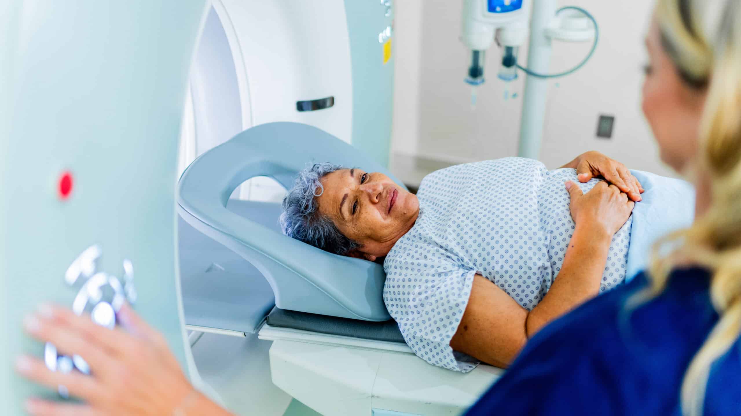
At Mountainside Medical Center, our cardiovascular imaging specialists provide comprehensive imaging tests to help prevent heart disease and heart attack. One such test is the CCTA + Heartflow Analysis. This non-invasive test uses AI technology to create a personalized 3D model of your coronary arteries and determine whether heart disease may be impacting blood flow to your heart. We also offer $99 calcium CT scoring, as well as electrocardiography. Early insights into your heart health can have lifelong impact–take the first step towards a healthier heart and schedule an appointment with our heart imaging specialists today!

Diagnostic heart screens we provide
Heart disease is the No. 1 killer of Americans. However, most people don’t know they have it until they have a heart attack or stroke. At Mountainside Medical Center, we are proud to offer the latest in diagnostic technology to screen for heart problems before you ever experience a heart attack or develop heart disease. We offer the following heart screens:
- Cardiac catheterization: During a cardiac catheterization test, a small catheter is inserted through an artery or vein and dye is injected into the heart to detect blockages and other heart problems.
- CCTA + Heartflow Analysis: This non-invasive test uses AI technology to create a personalized 3D model of your coronary arteries and determine whether heart disease may be impacting blood flow to your heart.
- Computerized tomography (CT) calcium scoring: This scan shows the presence, location and extent of calcified plaque in the walls of the coronary arteries. Plaque buildup narrows the arteries, restricts blood flow to the heart and ultimately can lead to heart attack.
- Electrocardiograms – Also known as an EKG or ECG, an electrocardiogram is a simple, painless test that checks for problems with the electrical activity of your heart. Electrocardiograms help determine the cause of symptoms of heart disease such as unexplained chest pain, irregular heart beat, dizziness, and shortness of breath. We offer two types of ECGs:
- Transesophageal echocardiograms: By passing a small, thin tube down the throat into the esophagus, physicians can assess the overall function of the heart’s chambers and valves. To reduce discomfort, the back of the patient’s throat is sprayed with a topical anesthetic sedative to help relax the patient. The patient’s heart rate, heart rhythm, breathing, and blood pressure are carefully monitored during the test. The study takes about 30 minutes and the pictures taken of the heart are recorded for later review. This test is often performed when the results from a standard echocardiogram are not sufficient, or when your doctor feels a closer look at your heart would provide a more accurate diagnosis.
- Transthoracic echocardiograms: This non-invasive test utilizes ultrasound waves to provide a quick and extremely accurate assessment of the overall health of the heart. It can provide a wealth of helpful information when searching for congenital heart, pericardial, and valvular diseases.
- Holter monitoring — A Holter monitor is a continuous recording of a patient’s ECG for 24 hours. It helps the physician correlate symptoms of dizziness, irregular heart rhythms (palpitations), or blackouts. It starts with applying ECG electrodes to the chest. Thin wires connect the electrodes to a small tape recorder, which is secured to the patient’s belt or slung over the shoulder and neck. The recorder is worn for 24 hours and the patient is encouraged to continue his or her daily activities. A diary or log is provided so that the patient can record activity and symptoms. The Holter monitor has an internal clock which stamps the time on the ECG strips. These can be used to correlate the heart rhythm with symptoms or complaints. After 24 hours, the Holter monitor is returned to the laboratory for analysis.
- Stress testing — This test shows how your heart works during physical stress, and is a useful screening tool for significant coronary artery disease. While connected to an electrocardiogram (ECG/EKG) machine, patients either walk on a treadmill or are given an intravenous (IV) medication that simulates exercise. The level of exercise is slowly increased and the patient’s symptoms and blood pressure response are consistently monitored throughout. The test continues until you reach a target heart rate, unless complications such as chest pain or an exaggerated rise in blood pressure develop. You will continue to be monitored for 10 – 15 minutes after exercising, or until your heart rate returns to baseline.
- Tilt table testing — This is a simple test that can help identify the causes of fainting by detecting a phenomenon known as postural or orthostatic hypotension. It involves placing a patient on a table with a foot-support, then tilting the table upward to a vertical position. The patient’s blood pressure, pulse, and symptoms are monitored throughout the test by a nurse and physician.
$99 CT Calcium Scoring
Heart screenings are crucial for identifying potential cardiovascular issues before they become serious or life-threatening. Many heart conditions, such as high blood pressure, arrhythmias, or blocked arteries, often develop without noticeable symptoms, making regular screenings vital for early detection. Early diagnosis through screenings allows for timely intervention, reducing the risk of heart attacks, strokes, and other complications. Additionally, heart screenings can provide insight into your overall health, empowering you to make informed lifestyle choices and work with your healthcare provider to prevent long-term damage.
At Mountainside Medical Center, we offer calcium-scoring tests to help you stay informed about your heart health. CT calcium scoring is a noninvasive test used to see inside your heart’s arteries and determine how much plaque has accumulated inside them. Arterial plaques are very common, and are typically made up of calcium, cholesterol, and fats that exist in the blood. CT calcium testing uses a computerized tomography (CT) scan to create a picture of your heart and arteries for your doctor, who can then work with you to create a plan of care that will work best for you.
Schedule your $99 heart screening today by calling 973-873-7787.
Prevent heart attacks with CCTA + HeartFlow Analysis
Coronary Artery Disease (CAD) is the most common type of heart disease and the leading cause of heart attacks. It occurs when plaque builds up in the coronary arteries, potentially restricting blood flow to the heart. This buildup can result in chest pain or even lead to a heart attack. Most people who suffer a heart attack don’t have prior symptoms and are considered low risk by traditional measures
A heart attack shouldn’t be the first sign of heart disease. CCTA + Heartflow Analysis offers a more comprehensive view of your heart’s health, allowing more informed decision making for you and your physician.
About CCTA + Heartflow Analysis
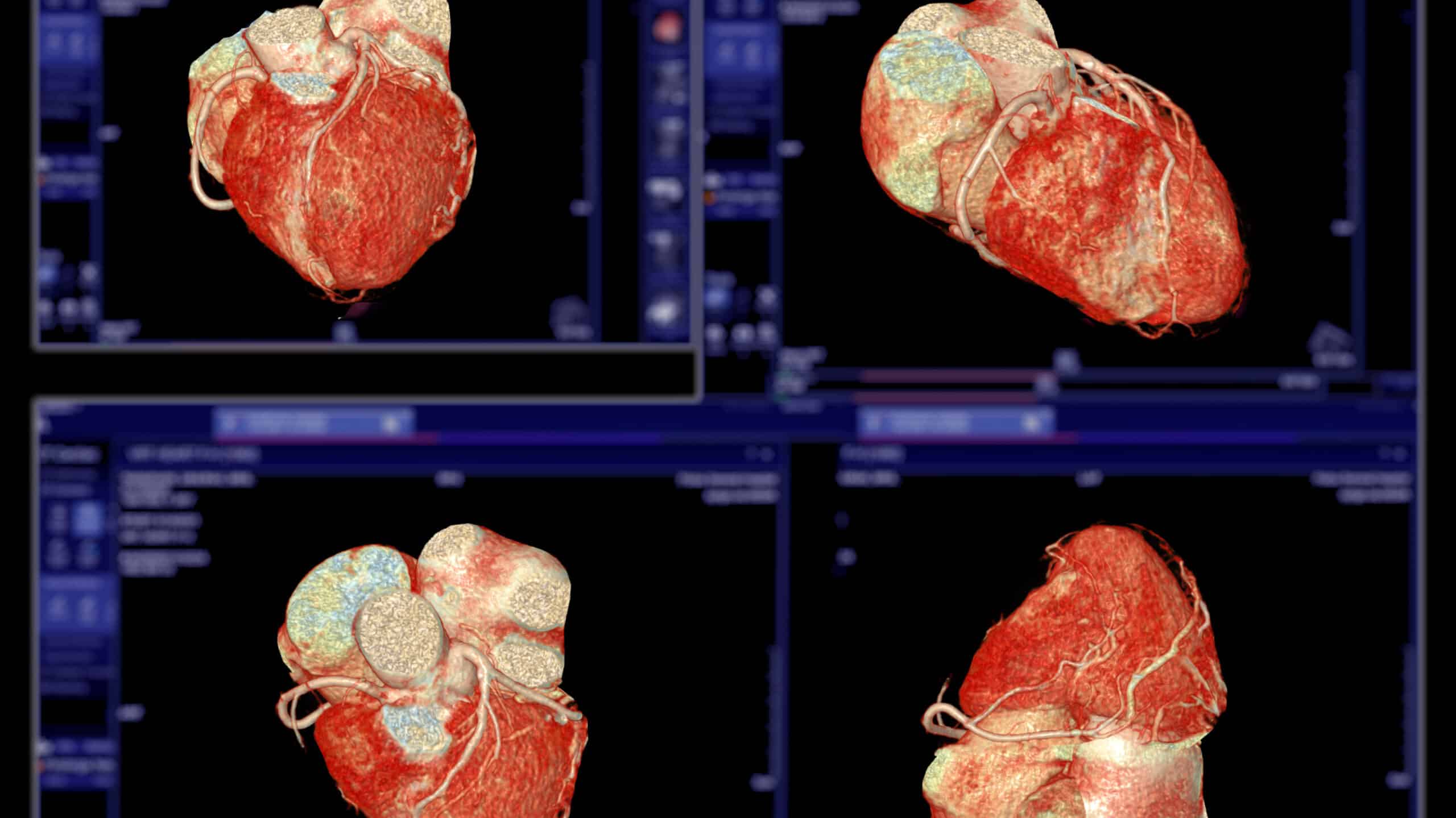
Mountainside Medical Center is advancing the assessment of your heart health with Heartflow Analysis. Using images from a non-invasive heart scan, also known as a coronary CTA, this AI-enabled heart test provides a personalized 3D model of your coronary arteries that shows how disease may be impacting blood flow to your heart, as well as identifying the amount, location, and type of plaque present.
This detailed information helps you and your doctor better understand the severity of your disease and determine a personalized treatment plan.
The benefits of CCTA + Heartflow Analysis
This innovative test offers many advantages. When you opt for this imaging test, you can expect:
- Accuracy: Heartflow Analysis has demonstrated superior accuracy in diagnosing CAD and is clinically proven to align with results from more invasive tests.
- A non-invasive procedure: Heartflow Analysis is a technology that uses images from your heart scan, so no invasive tests or anesthesia are needed.
- Personalized imaging: Heartflow builds a personalized, 3D model of your arteries, using images already taken during your heart scan.
- Comprehensive results: Heartflow Analysis can go beyond calcium scoring by identifying higher risk plaques that are most likely to cause a cardiac event.
- Guide for treatment decisions: Heartflow Analysis identifies how much and what type of plaque is present in your arteries and measures any blockages that could be limiting blood flow to your heart, providing information to help guide your personalized treatment plan.
How it works
Step 1: scan
Your doctor will order a non-invasive Coronary CTA (CCTA) scan of your heart to look for disease.
Step 2: Measure
Using your scan, the images undergo advanced AI processing to generate a personalized, 3D model of your arteries. Your report will measure blood flow and plaque buildup in your coronary arteries.
Step 3: Act
Your doctor receives a personalized, color-coded 3D model of your coronary arteries and detailed insights about your heart. With all the information in hand, you and your provider can make an informed choice on the best treatment pathway for you.

For more information, please call 973-873-7787.